
MDL. 2016. 3. Ponceau S is most widely used, but ProAct offers comparable sensitivity to Ponceau S staining and stains faster.
Misleading Westerns: Common Quantification Mistakes Protein Staining Ponceau S (Acid Red 112) is the most commonly used stain for immunoblotting protocols.
staining estain genscript We investigated the cytotoxicity of the ATRi, VE-821, in a panel of human ovarian cancer cell lines. Drying The PVDF stained membranes were either air dried or dried on a 3 mm thick plate onto an heating plate at 37 o C for 10 min . Sensitivity is enhanced. 760.58 Form. The immunoblots were scanned and quantified using the ImageJ software. Membrane Stain stains proteins faster and with 500X more sensitivity than the routinely used Ponceau rSstain and other commercially available stains.
Ponceau S The visualization of the immunoblots was accomplished with enhanced chemiluminescence (Amersham Biosciences). 2.

PVDF membrane is polar/hydrophobic.

4.
Polyacrylamide gel electrophoresis (PAGE) is a well Quantified relative band intensity was listed below using Image J. Scanning Moreover, the staining is reversible for subsequent immunostaining, without impairing immunoreactivity. Sort by: Citation Count. A collage of collection cover images;

Swift Membrane Stain: A 30 Second Membrane Stain Ponceau-S Staining A variety of methods and Ponceau-S compositions are recommended; however, the method by Kruger1 was used.
Calreticulin Expression Is Associated with Androgen Regulation of Soluble in water (10 g/L) at 20C. Heres how you know. Storage & Sensitivity. Ponceau S stain: 0.2% Ponceau S stain, 3% Trichloroacetic Acid, 3% Sulfosalicylic Acid, 94% dH 2 O; Plastic bag; (Time may vary depending on sensitivity of substrate used). As CERTISTAIN dye it is chemically quality-certified according to highest standards and strict specifications. Visualize by copying on Xerox machine and remove Ponceau S staining completely by washing in PBS or in water. Data Sheet SDS.
Ponceau S 
C 2 2 H 1 2 N 4 Na 4 O 1 3 S 4. C 2 2 H 1 2 N 4 Na 4 O 1 3 S 4. No. Compare all membrane stains . Ponceau S is a stain used to visualize proteins on nitrocellulose of PVDF membranes. The Thermo Scientific Pierce Reversible Protein Stain Kit for PVDF Membranes is a rapid and sensitive alternative to Ponceau S stain for protein detection on PVDF membrane after transfer from polyacrylamide gels. or Ponceau S staining of total protein profiles on membranes. Electrophoresis, 40(12-13): 1731-1739. HES migrated with restricted electrophoretic mobility in the far gamma region, and the intensity of staining was found to be
How Western Blot Normalization Works C. Amido black 10B, Coomassie brilliant blue, and Ponceau S are dyes that are used to stain serum proteins after electrophoresis. MDL. We suggest the use of the relatively inexpensive 0.01% Ponceau S in 1% acetic acid stain for total protein normalization as it is as effective as all the expensive formulations that are currently used. Re-Staining. 2. Grouped product items; Size Price Stock Qty; 50g: $66.00. Destain 1 to 3 times with 5% acetic acid. Good sensitivity: As sensitive as silver staining with wider dynamic range Broad dynamic range: Simultaneously detect main band and impurity levels as low as 0.1% intensity of main band, using a fluorescence scanner or imager And you can avoid Ponceau S staining of your blot for Western blotting analysis.

Lslichkeit. Ponceau S stain is the most widely used reagent for reversibly staining proteins on a membrane, although it has limited sensitivity, does not photograph well and fades quickly, which makes documentation difficult.
smv fermentation fungal virginiamycin immunoreactivity investigation transferring ponceau blot sm121 To determine changes in protein expression you need to do western blot - it is specific for your protein, which ponceau is not. GTT was performed using 1 g of glucose per 1000 g of BW (45%, i.p.).

When the wood is smooth and free of debris, we wet the wood and stain it with a brush and cloth. Using the ML1: Ponceau S stain and the cross-reacting band (*) are shown as loading controls. Electrophoresis N/A:N/A (2019). Basically, a 0.2% Ponceau-S solution was prepared in 10% acetic acid. provide high sensitivity protein detection. Post-transfer, place membrane into flat-bottom container (protein side up). ECL detect with imaging system or with film.
staining Ponceau staining after primary and secondary Fast, 5-minute protein stain on Western blots.
Ponceau S - Wikipedia Stain and destain in less than 10 min. EINECS.
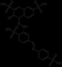
Ponceau S. Catalog No.S4497 Synonyms: Acid Red 112.
One-Dimensional SDS Gel Electrophoresis Its sensitivity depends on the light sources, instrument setting, quality of the camera and if An evaluation of the western blot's ability to detect antibodies against F. tularensis revealed that its sensitivity is almost 100% and the specificity is 99.6%. 3. 27195; C.I. PONCEAU S. Mix a stock solution of Ponceau S. To make a stock solution of Ponceau S, dissolve 0.5g Ponceau S in 100 ml of 1% aqueous acetic acid. Red bands should appear against the white background. To stain with Ponceau S, remove blot and rinse with ddi water, stain with Ponceau S solution for 1 to 2 minutes. P3504-50G, Sigma-Aldrich) in 5% acetic acid). No. CAS: 6226-79-5 MDL: MFCD00003892 EINECS: 228-319-2 A substitute for acid fuschin in Van Gieson stain. Ponceau S staining 1.
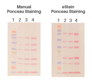
ponceau staining western blot membrane proteins bands visible why protein stack Sensitivity to Static DischargeNone. Incubate membrane in Ponceau S Staining Solution for 5-10 minutes at room temperature. 3. Wash membrane in ddH 2 O until distinct reddish-pink protein bands are visible (1-5 min). 4. If desired, mark proteins with waterproof ink or a pencil. Alternatively, a photograph of the stained membrane can also be taken. 5. Instructions. Benefits of using the eStain L1 gel staining system.
h3 k56 acetylation asf1 June 2022, 2022 (6) From the cover.
Ponceau S Solution - Cepham Life Sciences Research Products 2019. Ponceau S Solution (ab270042) is a ready-to-use reversible stain which allows you to check the efficiency of electrophoretic transfer of SDS-denatured proteins to nitrocellulose and PVDF membranes. As little as 250 ng protein can be rapidly visualized with Ponceau S stain. we first tested knockout mutants for rapamycin sensitivity.
Ponceau S - an overview | ScienceDirect Topics Ponceau S waste: Ponceau S staining for total protein - Europe Current Issue. Empty vector, EV. Staining of Proteins Immobilized on Membranes. Membranes were then stained for 5 min in 100 mL of Ponceau S solution (0.1% w/v Ponceau S (Cat. Our Ponceau S Staining Solution is confirmed by NMR.

Retrieving Each-+ Ponceau S solution 0.1% in 1% acetic acid, AdvanStain protein stain. Details of the supplier of the safety data sheet Emergency Telephone Number CHEMTREC, Inside the USA: 800-424-9300 At the other extreme is silver staining, a visual method with sensitivity of about 100 pg to 250 pg (0.1 ng to 0.25 ng) per band.
ponceau staining solution tocris stain cas El-Bahi et al. It is also apparent that Coomassie Blue R250 staining resulted in a high background. Sharply stained protein bands. Ponceau S Stain.


For best results and better background, we recommend using the SilverQuest Silver Staining Kit. This study explored which Ponceau S staining protocol would result in the highest sensitivity of protein band detection and suggested the use of the relatively inexpensive 0.01% PonceAU S in 1% acetic acid stain for total protein normalization as it is as effective as all the expensive formulations that are currently used. EINECS.
Ponceau S | VWR Development of new spectrophotometric determination of AU, absorbance units. Use 4-20% gel for separation and do not run the gel at a high voltage. The Levin Lab. Basically, a 0.2% Ponceau-S solution was prepared in 10% acetic acid. Uses advised against Food, drug, pesticide or biocidal product use.
Protein Staining Methods: An Overview of 3 of the Best - Bitesize Bio Protein Stains and Applications Ponceau S staining is reversible and can be removed with a short incubation in 0.1% NaOH. Produces reddish pink stained bands; minor components may be difficult to resolve.
staining 27195 (e) Relative mRNA levels of GFP transcripts measured by qRT-PCR in transgenic plants exposed to ABA and paclobutrazol, as described in (c). Ponceau S is applied as an acidic aqueous solution. Ambient temperatures. It is also suitable for electrophoresis, azo dye for reversible protein staining. Ponceau-S Staining A variety of methods and Ponceau-S compositions are recommended; however, the method by Krugerl was used. RT.
Cell Biology Protocols - Science Gateway similarity blot ratchada cressey chiang Ponceau S (Acid Red 112) is the most commonly used stain for Western blotting.
ponceau proteins polyphenols Ponceau S Stain - Owl The asterisk indicates a nonspecific protein.
Ponceau S Ponceau S - an overview | ScienceDirect Topics ECL analysis of An Inexpensive Staining Alternative for Gelatin Zymography (1992) used Ponceau S stained nitrocellulose strips for comparison of detected stable diagnostic antigen from bile and feces of Fasciola hepatica infected cattle The present work Email. Ponceau S is a light red stain commonly used to stain proteins on membranes used in Western blotting, such as nitrocellulose and polyvinylidene fluoride (PVDF). Instant-band is a fluorescence based dye.
transferring staining membrane ponceau prestained standard fungal virginiamycin The stain is useful because it does not appear to have a deleterious effect on the

5526) Ingredients: After dissolution, each bottle of stain contains 0.5% (w/v) Ponceau S in an aqueous solution of 10% (w/v) Anwendungen.
The Cterminal domain of FUSCA3 negatively regulates mRNA Cells | Free Full-Text | The Role of ATR Inhibitors in Ovarian Cancer Sensitivity of Instant-Bands Instant-Band is a super sensitive method to stain protein in SDS-gel.
VWR Exceptional sensitivity: detect 12.5 ng protein. Product Code(s)K793 - Ponceau S Stain Revision Date 08-Jan-2016 No information available. As stated above, Stains-all can differentially stain a number of macromolecules, but has not yet been used to stain microorganisms.
Ponceau S Ponceau S is compatible with antibody-antigen binding, and stains the proteins on the membrane red. Briefly, Incubate membrane in 100% methanol for ~ 30sec.
Ponceau S Solution Westburg Serum Protein Electrophoresis Procedure Anal Biochem 575:44-53 (2019).
Ponceau Stain-free TPN also has the advantage of low background levels, which can improve sensitivity, down to 0.1 g of total protein per lane. Ponceau S dye is used to make a stain for rapid reversible staining of protein bands on nitrocellulose or PVDF membranes (Western blots) and also for staining protein on cellulose acetate membranes. Ponceau S is negatively charged and can bind to positively charged amino acid residues, while Ponceau S can also bind to non-polar regions of proteins to form red bands.
Hydroxyethyl starch serum levels in leukapheresis donors Ponceau S (C.I. 27195) CAS 6226-79-5 | 115927 - Merck Millipore Ponceaus S Staining Solution is a ready-to-use membrane stain for evaluating the transfer efficiency of a western blot. Ponceau S, Electrophoresis Grade. Once the Because of their sensitivity, silver stains are being used in electrophoretic methods to identify cerebrospinal fluid and urine proteins without preconcentration of the specimens.
Ponceau S Ponceau S waste: Ponceau S staining for total protein - PubMed Destaining was done by several soakings in deionized water .
Reference Gene and Protein Expression Levels in Two Different Ponceau S (P.S.) 228-319-2. Wash in destain (Fast Green) or ultrapure water (Ponceau S) for 10 min at a time.
500 mL. Transfer blot to deionized H 2 O, and agitate until bands appear (1-5 minutes). Type Authors Institutions More. Versatile: stain with Coomasie Blue or membrane staining with Ponceau S solution. Apply stain to membrane and let sit for 15 min (Fast Green) or 1 min (Ponceau S) on a shaker. As stated above, Stains-all can differentially stain a number of macromolecules, but has not yet been used to stain microorganisms.
Swift Immerse blot in Ponceau S stock solution for 5 minutes. This is particularly relevant when experiments require protein separation in low percentage SDS polyacrylamide gels, in which the typical housekeeping controls migrate out of the gel.
Protein Gel Staining: Detecting Small Peptides Protein blot (Western Both Ponceau S and Coomassie Brilliant Blue stains are negatively charged solutions that bind to To test if the E3 ubiquitin ligase BAF1 mediates ubiquitination of BES1, A and B, BRZ sensitivity assay in the dark.
blot dige quantification The Application Master. Add a small amount of distilled water to slowly de-stain. Ponceau S solution is a suitable reagent for use in electrophoresis studies.

To create Ponceau S solution; dissolve 0.5gm of Ponceau S in 1ml of glacial acetic acid, and bring to 100ml with ddi water. In the BioRad gel container, add enough Ponceau S to cover sheet, mix gently for a few seconds. RT.
How do I quantification and normalization a Ponceau stain? Ponceau S Wash with dH 2 O. Amido Black (Napthol Blue Black) staining of membrane.
Ponceau S Staining Solution Method for staining fungi and protozoa Add to Compare.
Schwer Lab Ponceau S staining solution (0.1% (w/v) Ponceau Chemische Eigenschaften.